History of
Cataract Surgery
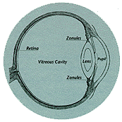
The term cataract refers to the clouding of the normally transparent crystalline
lens within the eye. The lens, located behind the pupil, is suspended in place by
thousands of tiny chemical strands called zonules. The lens focuses light upon the retina
at the back of the eye so that we can see clearly.

The word cataract comes from the Greek for waterfall. Until the mid 1700s, it
was thought that a cataract was formed by opaque material flowing, like a waterfall, into
the eye. We now know that the clouding of the lens usually occurs as a result of natural
aging processes, metabolic changes, injury, various forms of radiation, or toxic chemicals
or drugs. People with cataracts have blurred vision, making everyday activities such as
driving and reading difficult. Successful cataract surgery restores the ability to perform
these activities.

The earliest written reference to cataract surgery is found in Sanskrit manuscripts
dating from the 5th century BC. They are thought to have been written by the Hindu surgeon
Susruta. He practiced a type of cataract surgery known as couching or reclination, in
which the cataractous lens was displaced away from the pupil to lie in the vitreous cavity
in the back of the eye. This displacement of the lens enabled the patient to see better.
Vision, however, was still blurred because of the unavailability of corrective lenses. As
recently as the middle of this century, couching was still practiced in Egypt, India, and
Tibet.
In the Western world, recent excavations in Babylonia (Iraq), Greece, and Egypt have
uncovered bronze instruments that would have been appropriate for cataract surgery. The
first written description of the cataract and its treatment in the West appears in 29 AD
in De Medicinae, the work of the Latin encyclopedist Celsus. He describes in this
work the practice of needling, or discission, of cataracts, a technique in which the
cataract is broken up into smaller particles, thereby faciliting their absorption.
Interestingly, Hippocrates does not refer to cataract surgery in his writings. Galen,
the pivotal medical figure of antiquity whose theories went unchallenged for more than
1,500 years, erroneously believed that the lens rather than the retina was the seat of
vision, and that itsremoval would cause blindness. History also records the use of
bloodletting, antiphlogistics (agents to counteract inflammation and fever), and mercury
to prevent or dissolve cataracts - all of which were unsuccessful.
More advances continued to be made in lens removal. In 1957 Barraquer of Spain used
alpha-chymotrypsin to enzymatically dissolve the zonules for removal of the lens.
Cryo-surgery was introduced by Krawicz of Poland in 1961 to remove the lens with a tiny
probe that could attach by freezing a small area on the surface of the cataract.
Various forms of cataract aspiration were tried using curettes, hollow glass rods for
sucking the lens material out of the eye. In the late 1960s Charles Kelman of New York
developed a technique for emulsifying the lens contents using ultrasonic vibrations and
aspirating the emulsified cataract.
While the evolution of techniques for removing cataracts led to a very safe and
successful operation, it left the patient aphakic, meaning without a lens in the eye to
focus the image on the retina. The use of thick, heavy spectacles postoperatively was
somewhat helpful, and the later development of contact lenses provided even better
restoration of vision, although contact lenses could only be tolerated by a small
percentage of patients. It wasn't until Harold Ridley introduced the intraocular lens in
England in the 1940s that efficient and comfortable visual rehabilitation became possible
following cataract surgery. The intraocular lens, or IOL, is a permanent plastic lens
implanted inside the eye to replace the crystalline lens. In recent decades, there has
been a rapid evolution of designs, materials, and implantation techniques for intraocular
lenses, making them a safe and practical way to restore normal vision at the time of
surgery.

Modern cataract surgery, in which the cataract is actually extracted from the eye, was
introduced by Jacques Daviel in Paris in 1748. Daviel advocated a form of
extracapsular surgery in which the inner lens contents were removed from the eye, but a
portion of the lens capsule or outer covering and the zonules that attached it were left
in place. Samuel Sharp of London introduced the concept of intracapsular cataract surgery
in 1753 by using pressure with his thumb to remove the entire lens intact through an
incision. Small suction cups (erysiphakes) were introduced for this purpose in 1902 as
well as various capsular forceps to grasp the lens for removal.
The use of sutures for cataract surgery was first described by Henry Willard Williams
of Boston in 1867, using a shortened sewing needle threaded with a strand of fine glover's
silk. Manufacturing techniques have since led to the development of extremely fine but
strong sutures and very small, firm steel needles, which in conjunction with the
microscope in the operating room have led to major improvements in the safety and
long-term results of cataract surgery.
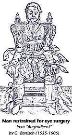
It wasn't until the 1840s that general anesthesia was introduced for surgical
procedures. Previously, the services of a strong assistant were required to hold the
patient's head still while the surgery was being performed. In 1884 anesthesia in the form
of eyedrops (cocaine) was developed, obviating the hazards of general anesthesia and its
postoperative complications. Cocaine has subsequently been replaced with other
anesthetics.
The techniques and materials historically developed for cataract surgery, in particular
the rapid improvements of recent decades, have made possible the miracle of modern
cataract surgery. Today, patients can have their cataracts safely removed as an outpatient
procedure, under local anesthesia, with the implantation of a sophisticated intraocular
lens calculated to correct their vision, and can resume their normal activities in a
matter of days.

What does the future hold? Improved knowledge of toxic chemicals, cataract-causing
drugs, and harmful radiation may enable physicians to reduce the incidence of cataracts.
Perhaps eventually an agent will be discovered that will slow or prevent the aging process
that causes the lens to become cloudy. There is reason to hope that such an agent will
eventually be identified and, if so, the need for cataract surgery will be significantly
reduced.
|